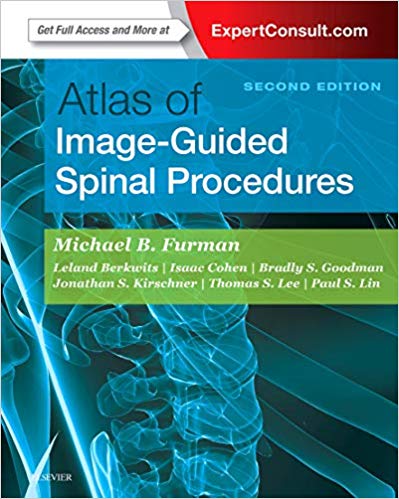
Atlas of Image-Guided Spinal Procedures – 2018
اطلس روشهای ستون فقرات با هدایت تصویر – ۲۰۱۸
نسخه دوم اطلس روشهای نخاعی با هدایت تصویر دارای یک قالب اطلس بسیار بصری است که دقیقاً نحوه انجام هر تکنیک را نشان می دهد. این مرجع پزشکی هر مرحله ، گام به گام شما را طی می کند تا با خیال راحت و کارآمد درد بیماران را تسکین دهید. این کتاب یک روش الگوریتمی ، هدایت کننده تصویر برای هر تکنیک ارائه می دهد. نمای مسیر (نشان دهنده “راه اندازی” فلوروسکوپی). نمایش تأیید چندخطی (AP ، جانبی ، مورب). و “نمای ایمنی” (آنچه باید هنگام تزریق از آن اجتناب شود) ، همراه با الگوهای کنتراست بهینه و زیر حد حداکثر. هر فصل فلوروسکوپی و سونوگرافی نیز همان “صدا” را دارد ، بنابراین می توان به راحتی دنبال کرد.
Weir & Abrahams’ Imaging Atlas of Human Anatomy
Product details
- Hardcover: ۶۸۰ pages
- Publisher: Elsevier; 2 edition (December 21, 2017)
- Language: English
- ISBN-10: ۰۳۲۳۴۰۱۵۳۸
- ISBN-13: ۹۷۸۰۳۲۳۴۰۱۵۳۱
The second edition of Atlas of Image-Guided Spinal Procedures features a highly visual atlas format to illustrate exactly how to perform each technique. This medical reference walks you through each procedure, step-by-step, to safely and efficiently relieve patients’ pain. This book presents an algorithmic, image-guided approach for each technique; trajectory view (demonstrates fluoroscopic “setup”); multiplanar confirmation views (AP, lateral, oblique); and “safety view” (what should be avoided during injection), along with optimal and suboptimal contrast patterns. Each fluoroscopic and ultrasound chapter also has the same “voice” so it is easy to follow.
- Safely and efficiently relieve your patients’ pain with consistent, easy-to-follow chapters that guide you through each technique.
- Presents an algorithmic, image-guided approach for each technique: trajectory view (demonstrates fluoroscopic “setup”); multiplanarconfirmation views (AP, lateral, oblique); and “safety view” (what should be avoided during injection), along with optimal and suboptimal contrast patterns.
- Special chapters on Needle Techniques, Procedural Safety, Fluoroscopic and Ultrasound Imaging Pearls, Radiation Safety, and L5-S1 Disc Access provide additional visual instruction.
- View drawings of radiopaque landmarks and key radiolucent anatomy that cannot be viewed fluoroscopically.
- Videos including procedural “set-up” and optimal and suboptimal constrast flow are available in the Expert Consult eBook version.
- Includes new unique and diagrams demonstrating cervical, thoracic and lumbar radiofrequency probe placement and treatment zones on multiplanar views, as well as new unique tables and examples differentiating between optimal and suboptimal epidural contrast flow
- Features new coverage of ultrasound techniques, as well as new presentation of procedures for “spine masqueraders” such as the hip and shoulder.
- Expert Consult eBook version included with purchase. This enhanced eBook experience allows you to access and search all of the text, figures, images, videos, and references from the book on a variety of devices
-
تخفیف %25 تخفیف
Diagnostic Pathology: Infectious Diseases 3rd Edition
۸,۹۵۰,۰۰۰ ریال – ۹,۳۵۰,۰۰۰ ریال -
تخفیف %26 تخفیف
Clinical Cardiac Electrophysiology: A Practical Guide
۶,۶۵۰,۰۰۰ ریال – ۷,۰۰۰,۰۰۰ ریال -
تخفیف %29 تخفیف
Foundations of Maternal-Newborn and Women’s Health Nursing
۱۲,۷۰۰,۰۰۰ ریال – ۱۳,۵۰۰,۰۰۰ ریال -
تخفیف %28 تخفیف
Textbook of Diagnostic Sonography: 2-Volume Set 9th Edition
۲۹,۰۰۰,۰۰۰ ریال – ۳۲,۰۰۰,۰۰۰ ریال -
تخفیف %29 تخفیف
Medicine Morning Report Subspecialties: Beyond the Pearls
۹,۹۵۰,۰۰۰ ریال – ۱۰,۹۵۰,۰۰۰ ریال

نقد و بررسیها
هنوز بررسیای ثبت نشده است.