Pocket Atlas of Sectional Anatomy , Vol. II: Thorax, Heart, Abdomen and Pelvis | 2014
تمام رنگی
وزیری
پروسه چاپ این کتاب بین 5 الی 7 روز کاری میباشد
اطلاعات بیشترقیمت منصفانه
ارسال سریع
تنوع و کیفیت بالا
پشتیبانی و پاسخگویی
-
%50 تخفیف

آناتومی سینه (توراکس) | جلد اول - چاپ سوم
قیمت اصلی: ۴,۰۰۰,۰۰۰ ریال بود.۲,۰۰۰,۰۰۰ ریالقیمت فعلی: ۲,۰۰۰,۰۰۰ ریال. -
%10 تخفیف

اطلس توصیفی آناتومی دورلند / گری
قیمت اصلی: ۱۲,۸۰۰,۰۰۰ ریال بود.۱۱,۵۰۰,۰۰۰ ریالقیمت فعلی: ۱۱,۵۰۰,۰۰۰ ریال. -
%18 تخفیف

کلیات آناتومی - دکتر مهدی زاده ( آناتومی عمومی + آزمون )
قیمت اصلی: ۴,۸۰۰,۰۰۰ ریال بود.۳,۹۵۰,۰۰۰ ریالقیمت فعلی: ۳,۹۵۰,۰۰۰ ریال. -
%20 تخفیف

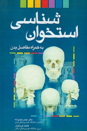
استخوان شناسی به همراه مفاصل بدن
قیمت اصلی: ۴,۰۰۰,۰۰۰ ریال بود.۳,۲۰۰,۰۰۰ ریالقیمت فعلی: ۳,۲۰۰,۰۰۰ ریال. -

Pocket Atlas of Human Anatomy
۵,۹۰۰,۰۰۰ ریال -

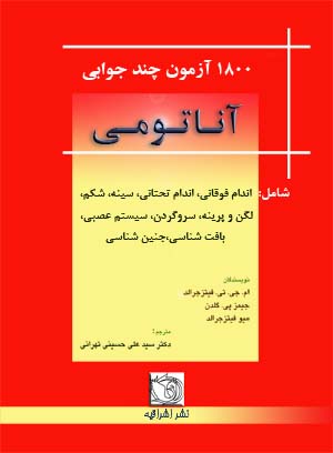
آناتومی- 1800 آزمون چند جوابی
۳,۵۰۰,۰۰۰ ریال

978-3131256041
ویراست چهارم
4
تمام رنگی
وزیری
346
Pocket Atlas of Sectional Anatomy , Vol. II: Thorax, Heart, Abdomen and Pelvis | 2014
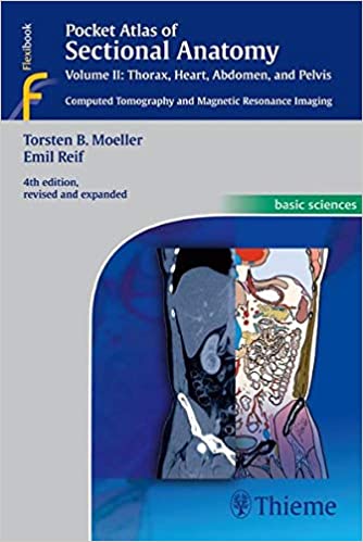
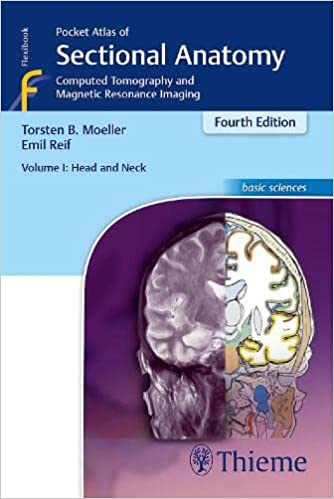
خرید کتاب افست اطلس آناتومی مقطعی : جلد دوم – توراکس قلب ابدومن و پلویس – ۲۰۱۴
Computed Tomography and Magnetic Resonance Imaging
This comprehensive, easy-to-consult pocket atlas is renowned for its superb illustrations and ability to depict sectional anatomy in every plane. Together with its two companion volumes, it provides a highly specialized navigational tool for all clinicians who need to master radiologic anatomy and accurately interpret CT and MR images.
Special features of Pocket Atlas of Sectional Anatomy:
- Didactic organization in two-page units, with high-quality radiographs on one side and brilliant, full-color diagrams on the other
- Hundreds of high-resolution CT and MR images made with the latest generation of scanners (e.g., 3T MRI, 64-slice CT)
- Color-coded schematic drawings that indicate the level of each section
- Consistent color coding, making it easy to identify similar structures across several slices
Updates for the 4th edition of Volume II:
- CT imaging of the chest and abdomen in all 3 planes: axial, sagittal, and coronal
- New back-cover foldout featuring pulmonary and hepatic segments and lymph node stations
- Follows standard international classifications of the American Heart Association for cardiac vessels and the AJCC/UICC for mediastinal lymph nodes
Compact, easy-to-use, highly visual, and designed for quick recall, this book is ideal for use in both the clinical and study settings.
Pocket Atlas of Sectional Anatomy : Vol. II |
Computed Tomography and Magnetic Resonance Imaging
This comprehensive, easy-to-consult pocket atlas is renowned for its superb illustrations and ability to depict sectional anatomy in every plane. Together with its two companion volumes, it provides a highly specialized navigational tool for all clinicians who need to master radiologic anatomy and accurately interpret CT and MR images.
Special features of “Pocket Atlas of Sectional Anatomy” Didactic organization in two-page units, with high-quality radiographs on one side and brilliant, full-color diagrams on the other Hundreds of high-resolution CT and MR images made with the latest generation of scanners (e.g., 3T MRI, 64-slice CT) Color-coded schematic drawings that indicate the level of each section Consistent color coding, making it easy to identify similar structures across several slices
Updates for the 4th edition of Volume II: CT imaging of the chest and abdomen in all 3 planes: axial, sagittal, and coronal New back-cover foldout featuring pulmonary and hepatic segments and lymph node stations Follows standard international classifications of the American Heart Association for cardiac vessels and the AJCC/UICC for mediastinal lymph nodes
Compact, easy-to-use, highly visual, and designed for quick recall, this book is ideal for use in both the clinical andstudy settings.
- Publisher : Thieme; 4th edition (September 1, 2013)
- Language: : English
- Paperback : ۳۴۶ pages
- ISBN-10 : ۳۱۳۱۲۵۶۰۴۴
- ISBN-13 : ۹۷۸-۳۱۳۱۲۵۶۰۴۱
- Item Weight : ۱ pounds
- Dimensions : ۵ x 0.8 x 7.5 inches
- Best Sellers Rank: #۱,۴۱۳,۲۹۳ in Books (See Top 100 in Books)
- #۸۳۷ in Anatomy (Books)
- #۹۲۹ in Physiology (Books)
- #۱,۴۴۵ in Medical Anatomy
-
%12 تخفیف

-
%12 تخفیف

-
%12 تخفیف
-
%25 تخفیف


دیدگاهها
هیچ دیدگاهی برای این محصول نوشته نشده است.